ATTN : Prof. Marta Brown, Dr. JAW Webb, Page 1 02/01/2020
Page 1 sur 21 Unpublished paper
Etienne-Jules Marey and Gaston Contremoulins, from Photography to Radiology : a centennial example of Translational Medicine.
Jean-François Moreau, MD, AIHP, FACR.
Emeritus Professor of Radiology, Université Paris Descartes
Consultant geriatric radiologist, Hôpital Corentin Celton, Issy-les-Moulineaux,
France.
Because of characters like Etienne-Jules Marey and his
photographer, Gaston Contremoulins, radiology has been a
science, an art, an industry and a business since its origin providing an
early example of translational technology to medicine and conversely.
Fig. 1. Four main characters (from top to bottom and left to right)
a) Etienne-Jules Marey in 1894 (Courtesy of Académie des sciences-Institut de France);
b) Gaston Contremoulins (Courtesy of Ecole de Radiologie de Saint-Germain-en-Laye);
c) Félix Guyon (Courtesy of Archives de l'AP-HP); d) Charles Rémy (author's collection).
Around the year 2000, meanwhile the
whole World just had concelebrated
the centennial of the discovery of X-rays by
Roentgen in 1895 (1), two new paradigms were
emerging almost simultaneously: «Disruptive
innovation» at the Harvard Business School
(2), and « Translational medicine » whose
most common definition was given by
the Dean of Stanford University, Philip A
Pizzo, in 2002 (3) : « From my perspective,
translational medicine can have both a
narrow as well as a more general definition.
Perhaps the most specific definition is
«bench-to-bedside» research wherein a basic
laboratory discovery becomes applicable
to the diagnosis, treatment or prevention of
a specific disease and is brought forth by
either a physician-scientist who works at the
interface between the research laboratory
and patient care or by a team of basic and
clinical science investigators. » An example
illustrating both of them is the sudden birth
and the quick growth of medical radiology in
Western Europe and the USA before World
War 1. This shows the performances of
humans facing innovations when they have
the power on hands either to develop or to
inhibit their applications to the patient's care
and cure with or without a long-term benefit
for the individuals and/or the populations.
Etienne-Jules Marey (1830-1904) belongs
to a group of French scientific personalities
who made important contributions to the
early growth of the new medical field induced
by the discovery of X-Rays (Fig.1). For the
whole of the twentieth century, their work has
been cast in shadow by the imperial character
of Antoine Béclère (4,5). There is no doubt
Antoine Béclère (1856-1939) is the official
father of French medical radiology since he
was convinced by the first demonstration
of the radiographic effect of X-rays that
Toussaint Barthélémy and Paul Oudin gave
in mid-January 1896 in their private office
close to the Hôpital Saint-Louis in Paris. Like
most of the pioneers, Béclère bought his first
equipment with his own money. He was the
first to teach radiology in the academic unit he
opened in 1899 at the Hôpital Saint-Antoine
in Paris and he wrote most of the medical
volume in the first French treatise dedicated
to radiology edited by Charles Bouchard and
published in 1904 (6). At the famous 1900
Exposition Universelle de Paris, he took the
opportunity to convene a hundred dedicated
individuals from all over the world to
participate at what was named the "Premier
Congrès International d'Electrologie et de
Radiologie Médicales" (7). They agreed
upon the creation of two words still officially
used to define a science, radiology, and
a job, radiographer. The latter split into
two kinds of subspecialists: the medical
doctors had become "radiologists" while
"radiographers" remained linked with the
practice of the technical work. The ubiquitous
corporatist conflict between both radiologists
and radiographers started in France in 1901.
Antoine Béclère was the head of the medical
camp that ultimately won the leadership, but
no earlier than 1935. The strongest resistant
radiographer, Gaston Contremoulins (1867-
1950), who founded the first laboratory
of radiology at the Hôpital Neckerin 1898
retired in 1934. Béclère was the third President
of the International Society of Radiology
(ISR) after the British Thurstan Holland and
Fig. 2. Béclère's stamp
(Courtesy of Centre Antoine Béclère).
the Swedish Gösta Forssell and before the
Swiss Hans Schinz and the American Arthur
Christie. There are a stamp (Fig. 2) and a
Hôpital Antoine Béclère opened in 1969 in
the city of Clamart, on a suburb of Paris. The
biennial Béclère Medal and Béclère Lecture
are the highest ISR awards granted with a
donation of his daughter, Antoinette Beclère,
to the ISR before she died in 1981.
And what about Etienne-Jules Marey?
Marey was elected in 1878 to the
Académie des Sciences, as the successor
of Claude Bernard (1813-1878). Both of
them were brillant medical doctors trained
at the Hospitals of L'Assistance publique à
Parisand are both sacred monsters of the
French physiology of the nineteenth century.
Bernard was mostly a chemist, Marey was
a physicist and an artist. Marey soon had a
passionate interest in the physiology of the
movement of the fluids and of living beings
(8). He was professor of physiology at the
Collège de France where he developed
chronophotography and pioneered the future
of cinematography with his assistant Georges
Demenÿ (9,10). Parallel work was undertaken
by Eadweard Muybridge, the photographer
of Leland Stanford, governor of the state of
California, USA, and founder of Stanford
University in Palo Alto (11). Both of them
worked together fruitfully when Muybridge
visited Marey's Station physiologique during
several months in 1881 (12). Marey was a
so prestigious scientist that he presided over
the centennial of the creation of the Institut
de France, encompassing the five greatest
National Academies including the Académie
des Sciences; that event was celebrated
during the notable year 1895, marked by the
foundation created by Alfred Nobel in Paris
and the discovery of X-rays. Marey used his
speech on 23 December 1895 at the end of his
mandate to express his sorrow over the recent
death of Louis Pasteur (13). He could not cite
the discovery of X-rays because Wilhelm
Roentgen did not disseminate the information
worldwide before 5 January 1896. Before
then only Roentgen's wife, whose hands
were imaged twice on the 9 November
and 22 December 1895, was aware of it ;
Roentgen informed the Wurzburg Physico-
Medical Society on 28 December 1895. The
mathematician Henri Poincaré was the only
Frenchman among the illustrious scientists
who received the personal letter and the
reprint Roentgen mailed on 2 January 1896
(1,4,7).
Poincaré and the medical membership
of the Académie des Sciences received
the news in January: Etienne-Jules Marey,
Charles Bouchard, Félix Guyon, Pierre-Carl
Potain, Arsène d'Arsonval, Odilon-Marc
Lannelongue and Paul Brouardel who was
the Dean of the Faculté de médecine de Paris
(14). All of them save Brouardel were early
supporters of radiology. On 20 January 1896
the Académie des Sciences was told about
the first French radiographic trial by Oudin
and Barthélémy (15) and their paper was
published by Lannelongue (16), a surgeon
who produced his own ones later (17,18). Dr.
Toussaint Barthélémy was Poincaré's nephew
but, as a subscriber, he learnt the news when
he red the famous Frankfurter Zeitung issue
dated on 7 January 1896 announcing the
sensational discovery: he spoke a fluent
German because he was born in Lorraine, a
province he quited in 1870 after the French
defeat versus the Prussian army; he informed
immediately his friend Oudin who was a
famous biophysicist; both of them repeated
Roentgen's experiments radiographing hands
successfully (4).
Auguste and Louis Lumière (19,20), Henri
Becquerel (21,22), Arsène d'Arsonval
(23) published their own observations on the
"photography through the opaque bodies".
The physicist Marie-Alfred Cornu was the
next president of the Académie des Sciences;
in his last speech on 21 November 1896
he put a great emphasis on the discovery
of X-rays and their medical applications as
the most important scientific event of the
year 1896 (24). One hundred and sixty-one
papers on Roentgen-rays or X-rays were
presented at the Académie des Sciences
during 1896 (25). Their number was meager
at the Académie Nationale de Médecine :
25 papers, all of them reporting medical
findings only (26). Ten out of these were
presented by Oudin and Barthélémy, most
of them illustrating the hand disease (27); all
of them were introduced by the professor of
dermatology and syphiligraphy at the hôpital
Saint-Louis, Alfred Fournier, who expressed
both his admiration for the procedure used
by the authors starting with the description
of the normal radioanatomy before they
described more and more radioclinical
patterns, and for the earnest improvement
of the quality of their images. Apart from a
few surgeons and the physicist d'Arsonval,
the earliest supportive medical academicians
were the bacteriologists; Béclère pionnered
virology and immunology before he became
a radiologist (5).
There is no doubt Marey was the catalyst
in promoting radiology through his
photographer, Gaston Contremoulins. In the
early 1890ies, the latter was appointed at
the laboratory of microphotography headed
by a trio made of Félix Guyon, professor of
urology at the Hôpital Necker, Mathias Duval,
professor of anatomy at the Ecole des Beaux-
Arts de Paris and of pathology at the Faculté
de médecine de Paris, and Yvon (4,10). More
precisely Contremoulins worked with Duval's
associate professor, Charles Rémy, who was
also both a surgeon at the hospital of the city of
Nanterre and a histologist at thehôpital de la
Charité(28). Who did inform Contremoulins
that Marey was developing microscopic
chronophotography? Guyon who was
Marey's close friend? More likely Georges
Demenÿ. In the fall 1892, Contremoulins
mailed a letter to Marey expressing his desire
to be his experimenter (10) (Fig. 3 ). After he
appointed Contremoulins under an unknown
agreement, Marey oversaw his experiments
on the microvasculature of small animals.
Marey and Demenÿ broke their long-term
worthwhile relationship in 1894 because of
a financial dispute dealing with the creation
the lucrative Société du Phonoscope by
the latter; Contremoulins did not take
over his position but he provided a strong
contribution to Delanglade's medical thesis
on chronophotography (29).
Like many photographers in the early
years of radiology (4), Contremoulins
rapidly became interested in the applications
of X-rays to medicine and biology. Félix
Guyon reported the first description of biliary
and renal calculi on 21 April 1896, at the
Académie Nationale de Médecine (30); the
experiment was done by James Chappuis,
professor of physics at the Ecole centrale des
Arts & Manufactures and a resident in urology,
Fernand-Joseph Chauvel, whom both were
also associated with the first radiography of
a fetus a few weeks earlier. Guyon quickly
bought X-ray equipment with his own money
and installed it in the Clinique Urologique
at the Hôpital Necker of Paris (31) where
he invited Contremopulins to perform his
radiographies (32). During the second half
of 1896, Marey presented two radiological
papers by Rémy and Contremoulins (33,34).
Marey persuaded them to study the vessels of
the hand with a radiopaque compound made of
metallic powders, generically termed bronze,
embedded in sealing wax then dissolved
in alcohol (Fig. 4). In 1897, both of them
published a method of radiophotography of
the soft tissues inspired by the histological
Fig. 4. Excerpt of Rémy and Contremoulins' first communication at the Académie des Sciences evidencing
the interest of Marey in the early development of radiology in 1896. (Facsimile by courtesy of Académie
des Sciences-Institut de France)
Fig. 3. Excerpt of Marey's letter dated on 14 December 1892 and mailed from Napoli, Italy, recommending
Contremoulins to his assistant Georges Demenÿ (Courtesy of Cinémathèque Française) : «Mr.
Contremoulins writes to me that he is highly motivated to working with you in photography. Before I'm back
[to Paris] then I show him what I'm doing in microscopic photography, let you do for him what you feel it is
convenient.» (Author's translation)
preparations by silver chromate (35).
Influenced by a report by Bouchard,
Dr. Georges Clemenceau, Ministre de
l'Intérieurwho also covered health affairs,
decided in 1898 to create four laboratories of
radiology in Paris (4). One of these, actually
dated on 1896, was already functioning under
the direction of Albert Londe, photographer of
the famous neurologist Charcot at the Hospice
de la Salpêtrière. According to a Wikipedia
anonymous resource (36), Albert Londe
with Marey performed many photographic
experiments concerning movement, and the
layout of his laboratory at the Salpêtrièrewas
similar to Marey's Station Physiologique.
In 1893, Londe published the first book on
medical photography, titled « La photographie
médicale: Application aux sciences
médicales et physiologiques ». In 1898, he
published « Traité pratique de radiographie
et de radioscopie: technique et applications
médicales» that is likely the first of the
discipline. Another laboratory was created
at the Hôpital Necker under the exclusive
Fig. 5. Contremoulins' compass (Courtesy of Ecole
de radiologie, Saint-Germain-en-Laye).
Fig. 6. Spectro-trigono-métro-radiographeused for
the localisation of intracranial foreign body. Drawing
published in L'Illustration, 22 November 1897
(Courtesy of L'Illustration).
Fig. 7. Monthyon-awarded Contremoulins' letter
acknowledging the Académie des Sciences written
on Station Physiologique stationery. (Courtesy
of Académie des sciences-Institut de France).
executive direction of Gaston Contremoulins
(4). From 1898 to 1901, Contremoulins built
an outstanding laboratory subsidized by the
city of Paris which became the reference for
the further projects in radiological wards.
He was not medically qualified but soon he
developed excellent relationships with the
surgeons who appreciated his meticulous
performance and his inventive abilities shown
in the idea of "métroradiologie" (Fig. 5).
With Rémy he had become famous in 1897
with a description of some sophisticated
and precise equipment for the detection of
foreign bodies they called "spectro-trigonométro-
radiographe" (37) ; they received
the Prix Monthyon a high medical award
delivered annually by the Académie des
Sciencesto compensate the expansive cost
of the tool (2500francs to be compared with
the 2000francs of Demenÿ's annual salary).
Contremoulins soon became a contributor to
journals and newspapers. The popular weekly
magazine, L'Illustration, dedicated the cover
page of the 22 November 1897 issue to that
innovative tool (Fig. 6 and 7). Another Prix
Monthyonwas awarded for his paper on the
technique of plain film used for the detection
of urolithiasis in humans published in 1899
with the urologist Albarran (38). Marey
presented several Contremoulins' papers
when he was the president of the Académie
Nationale de Médecine during the year 1900.
Contremoulins no longer communicated
with the Académie de Médecine afterwards,
because in 1901 he had become Béclère's
toughest enemy in a severe corporatist
conflict (4,5,32).
Antoine Béclère had to face opposition
both from a medical and a radiographic
lobby over four decades:
1) Béclère could not get the Ministère de
l'Educationto create an academic chair of
radiology before he died in 1939 (5). Many
medical academic colleagues were angry with
him because they believed he was perverting
the art of medicine, even though he respected
Laennec's anatomoclinical principles
and method (39). Still in the 1930ies,
they regarded radiology, a photographic
technology, with contempt. They disliked the
facts that radiology could rapidly provide a
precise diagnosis which might contradict
their intellectual arguments and that autopsy
was no longer the only means to make a
diagnosis anatomically. These disagreements
were topics for caricaturists until medicine
became truly scientific in the second half of
the 20th century.
2) Béclère also came into conflict with the
non medical radiographers, even when they
were respected, like Contremoulins, because
of the many scandals relating to incompetent
or dishonest practitioners. When a new deal
emerged in 1908 still under Dr. Georges
Clemenceau's Ministership, Béclère could
not obtain the chairmanship of the radiology
department exclusively for medical doctors
although this was recommended by the
Académie Nationale de Médecine (4,5).
Thirty-nine members of the Académie
des Sciences, headed by Paul Villard who
discovered the gamma-rays, signed a letter
supporting Contremoulins after they visited
his laboratory (4). However, neither Bouchard
nor Becquerel, who were respectively
Présidentand Secrétaire perpétuel of the
Académie des Sciences in 1909, nor the
medical membership with the exception of
the bacteriologist Emile Roux, co-signed the
letter. Contremoulins at the Hôpital Necker
and Puthomme, his former assistant, at the
Salpêtrière, kept their positions as the heads of
those two out of the eight official laboratories
of the hospitals of the Assistance publique à
Pariscreated by Clemenceau. Béclère was in
charge of the radiological service of the city of
Paris during World War1 (4,5). Contremoulins
refused to be under the hierarchy of a doctor
and resigned, in spite of his recognized
competence in the military aspects of the
work. Looking for foreign bodies was a
common radiological investigation. An
ad hoc committee was nominated in 1917
at the Académie de Médecine to evaluate
Contremoulins' method since it appeared that
the radiographer might have been unfairly
denigrated or ignored; Edouard Kirmisson
and three other academicians recalled the
interest in Contremoulins' metroradiological
tool in surgery (40).
Radiology would not have developed
without the convergence of three major
preliminary inventions : the Crookes' tube,
electrical power and photography. In 1901,
Roentgen was the first Nobel Prize winner
in physics, an honor that his compatriot
Phillipp Lenart did not obtain because, unlike
Röntgen, he was not a photographer then
could not evidence his own but controversial
discoveries (1,7). Contremoulins had all three
inventions at hand: he was able to make his own
Crookes'tubes, he knew electrical technology,
and he was an expert in photography. At the
Ecole des Beaux-Arts in Rouen, he learnt
the basics of multimodality imaging from
drawing to architecture. Working with Marey,
who was also a painter and a sculptor, he
became an inventive master of mechanics and
microscopic chronophotography. Working
with Rémy at the histology laboratory,
he learnt gross pathology in humans and
animals. Those who have had the chance to
look at his radiographs admire their superb
quality. His papers show his outstanding
mastery of both technology and diagnosis.
He had the privilege of radiographing the first
opacification of the subarachnoidal space with
Lipiodol by Forestier and Sicard in 1921 (41)
(Fig. 8). Contremoulins soon carefully studied
dosimetry and radioprotection (42,43,44). He
fought against the abusive use of fluoroscopy
and radiography. Many radiologists died
from radiation exposure or suffered terrible
radionecroses. Their names are listed in
a memorial in Hamburg, Germany, in the
hospital where Albers-Schoenberg practiced
radiology, but then died prematurely of
cancer (7). Many pioneers, like his colleague
Charles Infroit, successor of Albert Londe
who resigned in 1900, died prematurely
because they submitted themselves to
careless irradiations by the "invisible light"
before its harmful effects were clearly shown
at the 1910 Exposition Universelle of Liège,
Belgium that hosted the second «Congrès
International d'Electrologie et de Radiologie
Médicales». Neither Contremoulins nor his
staff members suffered any radionecrosis,
cancer or leukaemia; however they never
practiced radiation therapy. In the 1920s,
he wrote about the necessity to protect the
environment of the radiological wards, in
hospitals as well as in private offices (45,46).
In 1929, Maurice de Broglie, physicist and
expert in X-rays at the Académie des Sciences,
was in charge of an official report which
supported the validity of Contremoulins'
ideas about radioprotection in the structure of
radiographic rooms (47).
Marey as well as Roentgen were idealist
scientist contempting financial business.
Marey, who owned personal wealth, and
Demenÿ patented many inventions but they
were poor businessmen (10). Under the
auspices of the Collège de France, Marey
built and managed his Station physiologique
mostly with governmental subventions and
his own money, unlike the modern joint
ventures between research centers and
bankable start-ups. He invested 6000 francs
in six chronophotographs but he sold only one
specimen. In their correspondence exchanged
when Marey was used to spend the fall and
the winter seasons in Napoli, Italy, Demenÿ
always complains of financial problems
that his boss never tried to solve actually.
In spite of Demenÿ's pressure, Marey was
Fig. 8. First case of opaque myelography
after injection of Lipiodol in the subarachnoidal
space in 1921. Dr. Jacques Forestier
handwrote comments. Contremoulins
signed the radiography (arrow). (Courtesy
of Dr Michel Guerbet).
fully reluctant to involve his genious talent
in the development of cinematographic
entertainment programs that would have
subsidized their undertakings. Meanwhile
Demenÿ and his Société du Phonoscope fell
into bankruptcy in 1895, those programs
enriched talented "plagiarists" like Gaumont
and Lumière Brothers in France (48). Roentgen
Fig. 9 a, b, c. Metroradiological table and devices for radiopelvimetry detecting dystocies.
(Courtesy of Ecole de radiologie, Saint-Germain-en-Laye).
was even stricter since he refused to patent
the discovery of X-rays and to enter any
further business ventures. At an economical
viewpoint, radiology had been a disruptive
innovation that could develop free of charge
to all inventors of the equipments enabling a
new highly lucrative process of translational
technology to medicine. Contremoulins
who patented his invents as well as charged
the radiographies to the patients was wiser
at the Hôpital Necker where his laboratory
management never sounded to be negative
financially during his 35-year mandate.
Was Contremoulins an extraordinary
character? Studying his life and work,
the author's answer is : yes, indeed! Looking
at his radiopelvimetric method and tools
(Fig. 9 a, b, c), why not to evoke his inventive
potentials if he had lived when computed
imaging emerged from the 1960ies? His
expertise overwhelmed the strict boundaries
of radiology. With the surgeons Delbet,
then Schwartz and Robineau, he invented
the bone prostheses applied to numerous
wounded soldiers after World War 1 (49,50).
He invented many tools for orthopaedic
surgery (Fig. 10 and 11 ), which were usually
presented by Robineau at both Académies des
Sciences and Académie de Chirurgie (51,52).
Maurice Robineau (Fig.12) took advantage of
Fig. 10. Metroradiological device for osteosynthesis
of the knee. (Courtesy of Ecole de radiologie, Saint-
Germain-en-Laye).
Fig. 11. Prothesis of the cubitus made of aluminium
covered by latex. (Courtesy of Ecole de radiologie,
Saint-Germain-en-Laye).
Fig. 12. Dr Maurice Robineau. (Author's collection).
Contremoulins's inventiveness to propose the
first treatment (osteosynthesis) for fractures
of the neck of the femur by bone pinning in
France (53). In 1935, Contremoulins settled
in the city of Saint-Germain-en-Laye where
he continued to practice metroradiology at
the hospital. He founded a school there where
he taught future radiographers and this school
is still in operation. He committed suicide in
1950 because he had become blind (32).
In conclusion, because Etienne-Jules
Marey was in charge of eminent academic
positions while radiology was starting, he
had been immediately supportive of the
investment of his assistant photographer,
Gaston Contremoulins, in the promising
radiological specialty. He practiced at the
Hôpital Necker of Paris then at the Hôpital
de Saint-Germain-en-Laye during 54 years
of his life without any radio-induced damage.
Contremoulins was the radiographers' most
brilliant godfather. While he was in conflict
as early as 1901with the medical concept
of radiology headed by Antoine Béclère,
Contremoulins always found a strong group
of supporters at the Académie des Sciences
who would be active and faithful until he
retired in 1934 from the Hôpital Necker.
Both Marey and Contremoulins belongs to
the group of the ancestors of translational
medicine. Because Contremoumlins was
banned off the medical decision makers
before World War 2, the University of Paris
missed its chance to consolidate the core of
a future radiological institute of technology
where computed tomography programs could
have been conceived. His laboratory was
destroyed after he retired. The author was the
chairman of the Necker's new department of
radiology in 1988-99. He rebuilt it without any
knowledge of that prestigious but forgotten
story. Should he had known it, instead of
the destruction of a joint space unusable for
clinical radiology, he would have featured
the department differently with a prospective
vision of a technological laboratory to the
21st century when molecular radiology has
become the must of medical imaging.
Acknowledgement.
The author is highly indebted to Professor
Marta Braun, Ryerson University, Toronto,
Ontario, Canada, and to Dr. Judith A. W.
Webb, F.R.C.P., F.R.C.R., London, UK, for
their help in the edition of the manuscript.
The author thanks gratefully M.J.
Watremez and J.C. Stoleric who accepted
to communicate their unpublished memoir
written in 1983: «Gaston Contremoulins:
un pionnier de la Radiologie.» available at
the Ecole de Radiologie, Saint-Germain-en-
Laye, only.
The author gratefully acknowledges Mrs.
Claire Guttinger of the Department of
Archives of the Collège de France of Paris,
Mrs. Florence Greffe of the Department of
Archives of the Académie des Sciences, Mr.
Jean-François Vincent of the Bibliothèque
InterUniversitaire de Médecine (BIUM),
Mr. Valdo Kneubühler of the Cinémathèque
Française, and Mr. Laurent Provost of www.
Historix.fr website for their fruitful help in
the bibliographic research.
REFERENCES
1. R. A. Gagliardi, B. L. McClennan, Eds.
A History of the Radiological Sciences.
Diagnosis(Radiology Centennial, Inc.,
Reston, 1996).
2. J. L. Bower, C. M. Christensen,
Disruptive Technologies: Catching
the Wave. Harvard Business Review,
January-February 1995, pp1-13.
3. P. A. Pizzo, Letter from the Dean.
Stanford Medicine Magazine 19, 1
(2002).
4. G. Pallardy, M. J. Pallardy, A. Wackenheim,
Histoire illustrée de la Radiologie (Editions
Roger Dacosta, Paris, 1989).
5. A. Béclère, Antoine Béclère. (J.B.
Baillière, Paris, 1973).
6. A. Béclère, in C. Bouchard, Ed. Traité de
Radiologie Médicale (G Steinhel, Paris,
1904), vol 2.
7. E. R. N. Grigg, The trail of the invisible
light(Charles C Thomas, Springfield,
1965).
8. M.E. Silverman, Etienne-Jules Marey:
19th century cardiovascular physiologist
and inventor of cinematography. Clin.
Cardiol.,19, 339-341 (1996).
9. M. Braun, Picturing Time. The work
of Étienne-Jules Marey (1830-1904)
(University of Chicago Press, Chicago,
1992).
10. T. Lefebvre, T. Malthête, L. Mannoni,
Lettres d'Etienne-Jules Marey - Georges
Demenÿ, 1880-1894. (AFRHC /
Bibliothèque du film, Paris, 2000).
11. R. Solnit, River of Shadows : Eadweard
Muybridge and the Technological Wild
West(Viking Penguin, New York, 2003).
12. T. Lefebvre, Marey and
chronophotography (http://www.bium.
univ-paris5.fr/histmed/medica/marey/
marey03a.htm, Nov 2005)
13. E.J. Marey, Discours du président.
Séance publique annuelle du lundi 23
décembre 1895. C.R. Acad. Sci. 121, 1-14
(1895).
14. M.A. Cornu, Etat de l'Académie des
sciences. C.R. Acad. Sci. 122, 7-8 (1896).
15. P. Oudin, T. Barthélémy, Photographie
des os de la main, obtenue avec l'aide
des "X-Strahlen" de M le Professeur
Röntgen. C.R. Acad. Sci. 122, 150
(1896).
16. O. M. Lannelongue, P. Oudin,
T. Barthélémy, De l'utilité des
photographies par les rayons X dans
la pathologie humaine. C.R. Acad. Sci.
122:159-160 (1896).
17. O. M. Lannelongue, P. Oudin, Sur
l'application des rayons de Röntgen au
diagnostic chirurgical. C.R. Acad. Sci.
122, 283-285(1896).
18. O.M. Lannelongue, Application des
rayons X au diagnostic des maladies
chirurgicales. C.R. Acad. Sci.122, 695-
697 (1896).
19. A. Lumière, L. Lumière, Recherches
photographiques sur les rayons de
Röntgen. C.R. Acad. Sci. 122, 382-384
(1896).
20. A. Lumière, L. Lumière, A propos de la
photographie à travers les corps opaques.
C.R. Acad. Sci. 122, 463-465 (1896).
21. H. Becquerel, Sur les radiations émises
par phosphorescence. C.R. Acad. Sci.
122, 420-421 (1896).
22. H. Becquerel, Sur les radiations invisibles
émises par les corps phosphorescents.
C.R. Acad. Sci. 122, 501-503 (1896).
23. A. d'Arsonval, Observations au sujet de
la photographie des corps opaques. C.R.
Acad. Sci. 122, 500-501 (1896).
24. M.A. Cornu, Allocution lors de la séance
publique annuelle du lundi 21 décembre
1896. C.R. Acad. Sci. 123, 1099-1107
(1896).
25. M. Berthelot, Table des matières. C.R.
Acad. Sci. 122 1594-1598 and 123, 1357-
1358 (1896).
26. Radiographies. Table des Matières. Bull.
Acad. Nat. Med. 36, 936 (1896).
27. P. Oudin, T. Barthélémy, Table des
Matières. Bull. Acad. Nat. Med. 36, 932
(1896).
28. G. Huguet, Les professeurs de la faculté
de médecine de Paris. Dictionnaire
biographique 1794-1939. (Institut
national de recherche pédagogique &
Editions du CNRS, Paris, 1991) pp617-
618.
29. E. Delanglade, thesis, Faculté de
médecine de Paris, (1896).
30. F. Guyon, J. Chappuis, F.J. Chauvel,
Calculs rénaux et biliaires. Bull. Acad.
Nat. Méd. 35, 409-411 (1896).
31. P. Léger, Chroniques de l'Urologie
Française (Fondation Schering, Lys-les
Lannois, 1998) vol 1, pp67-103.
32. M.J. Watremez, unpublished material.
33. C. Rémy, G. Contremoulins, Endographie
crânienne au moyen des rayons Röntgen.
C.R. Acad. Sci. 123, 233 (1896).
34. C. Rémy, G. Contremoulins,
Emploi des rayons X pour les
recherches anatomiques: angéologie,
développements, ossifications, évolution
des dents, etc. C.R. Acad. Sci. 123, 711-
712 (1896).
35. C. Rémy, G Contremoulin, De la
radiophotographie des parties molles de
l'homme et des animaux. C.R. Acad. Sci.
124, 229-230 (1897).
36. Anonymous, A. Londe, http://
en.wikipedia.org/wiki/Albert_Londe, (3
December 2010).
37. C. Rémy, G Contremoulins, Appareil
destiné à déterminer d'une manière
précise, au moyen des rayons X, la
position des projectiles dans le crâne. E.J.
Marey, Discussion. C.R. Acad. Sci. 125,
831-836 (1897).
38. J. Albarran, G. Contremoulins,
Radiographie des calculs du rein. C.R.
Acad. Sci. 129:175-177 (1899).
39. B. Vergez-Chaignon, Les Internes des
hôpitaux de Paris. 1802-1952 (Hachette,
Paris, 2002).
40. E. Kirmisson, De l'emploi du compas
Contremoulins réglé d'après une
localisation radioscopique utilisant un
rayon normal vertical. Routier, Delorme,
Kirmisson, Reynier, Discussion. Bull.
Acad. Nat. Méd. 77, 522-526 (1917).
41. J. Arlet, Jacques Forestier, des stades aux
thermes. (Privat, Toulouse, 1988).
42. G. Contremoulins, Recherche d'une unité
de mesure pour la force de pénétration
des rayons X et pour leur quantité. C.R.
Acad. Nat. 134, 649-651 (1902).
43. G. Contremoulins, Appareil des
mesure des facteurs pénétration et
quantité de rayons X et totalisateur
radiophotométrique. C.R. Acad. Sci. 141,
26-29 (1905).
44. G. Contremoulins, E. Puthomme, De la
détermination des temps de pose. C.R.
Acad. Sci. 171, 383-1384 (1920).
45. G. Contremoulins, E. Puthomme, A
propos de la protection des tiers contre
les rayons X. C.R. Acad. Sci. 172, 1090
(1921).
46. G. Contremoulins, Le danger des rayons
X pour les voisins n'est pas un mythe. Le
Siècle médical. 60, 1 (1929).
47. M. de Broglie, Le danger des rayons X.
Le Matin. 23 septembre 1929.
48. L. Mannoni, Les premières années
de la société Léon Gaumont et Cie -
Correspondance commerciale de Léon
Gaumont, 1895-1899. (Afrhc , Paris,
1998).
49. G. Contremoulins, Sur le rôle de la
métroradiographie dans l'établissement
des pièces endoprothétiques en os mort.
C.R. Acad. Sci. 173, 1173-1176 (1921).
50. P. Delbet, M. Girode, G. Contremoulins,
Endoprothèse en caoutchouc armé pour
perte de substance du squelette. Bull.
Acad. Nat. Méd. 82, 110-111 (1919).
51. M. Robineau, G. Contremoulins,
Réactions de l'organisme humain sur les
pièces prothétiques et synthétiques en os
hétérogène stérilisé à l'alcool bouillant.
C.R. Acad. Sci. 180, 1543-1544 ().
52. M. Robineau, G. Contremoulins,
Exemples de synthèses et de prothèses
en os métal nu ou caoutchouté établies
sur des données métroradiologiques.
Résultats éloignés. C.R. Acad. Sci. 188,
1575-1576 (1929).
53. M. Robineau, G. Contremoulins,
Ostéosynthèse des fractures récentes
transcervicales du col du fémur. Presse
méd. 31, 825 (1923).
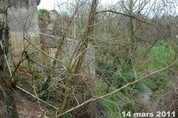
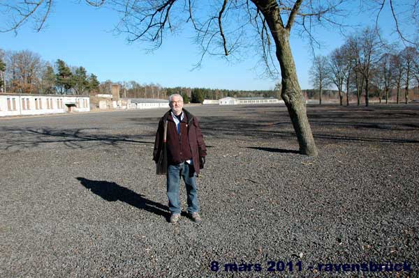
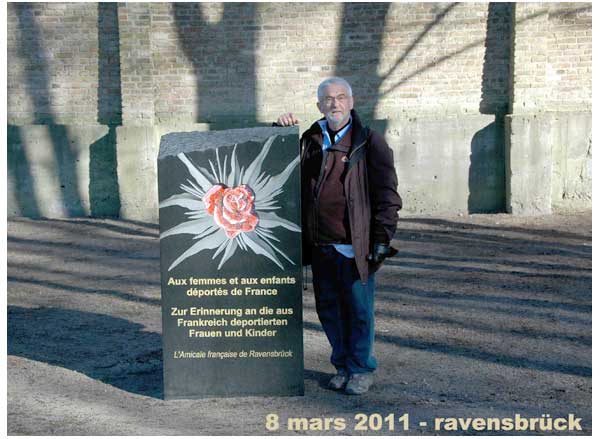
A RAVENSBRÜCK
LA PHARMACIE DE MARGUERITTE CHABIRON
A VERDELAIS ETAIT DANS CET IMMEUBLE
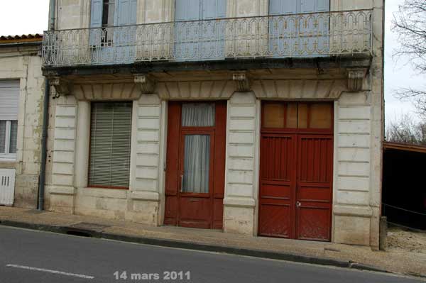
LES RESISTANTES S'ENFUIRENT PAR LE JARDIN A PIC